Physiology
- Contents of the abdomen are predominantly non-compressible fluid
- The pressure increases with inspiration and decreases with expiration
- A normal person has either a negative intra-abdominal pressure, or something in the range of 0-5 mmHg
- The morbidly obese and pregnant patient may have pressures as high as 10-15 mmHg
- In normal critically ill patients the pressure is 5-7 mmHg and the upper limit of normal is 12 mmHg
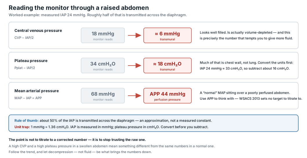
- Abdominal perfusion pressure is given by:
- Abdominal pressures > 35 mmHg result in compromised capillary perfusion of the intestine and bacterial translocation
- A target abdominal perfusion pressure ≥ 60 mmHg has been demonstrated to improve survival
Measuring Intra-Abdominal Pressure
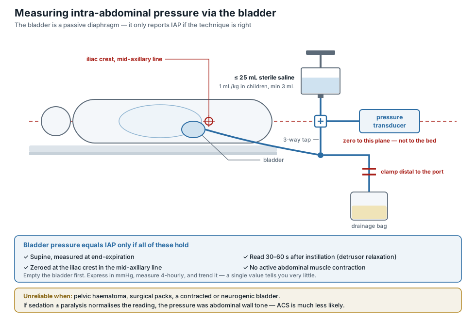
- The intra-abdominal pressure equals the bladder pressure when the following criteria are fulfilled:
- End of expiration
- Supine
- Zeroed at the iliac crest in the mid-axillary line
- Instilation of 25 mL of saline into the bladder (1 mL/kg for children up to 25 kg, minimum of 3 mL)
- Measured 60 seconds after instillation to allow detrusor muscle relaxation
- In absence of active abdominal muscle contraction
- Usually measured every 4 hours
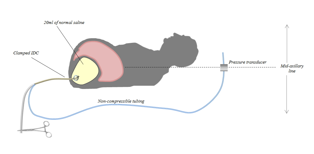
- A transducer set is attached to a side-port of the IDC
- The bladder is emptied completely then clamped
- 20 mL of saline is introduced into the bladder and after giving the detrusor 60 seconds to relax, the reading in the transducer is the intra-abdominal pressure
- Caveats
- Pressures may vary across individuals (with obese patients having higher baseline pressure values)
- As with all physiological phenomena, pressure gradients are more important than absolute pressure values
- Consequently, the intra-abdominal pressure value in isolation is inadequate to define abdominal compartment syndrome
- Pressure measurements may be inaccurate in the context of pelvic pathology (e.g., hematoma directly compressing the bladder)
- Interpretation among patients who are not intubated and breathing passively on mechanical ventilation is challenging
- If analgesia/sedation causes normalization of the intra-abdominal pressure, this makes abdominal compartment syndrome less likely
- If analgesia/sedation causes normalization of the intra-abdominal pressure, this makes abdominal compartment syndrome less likely
Definitions
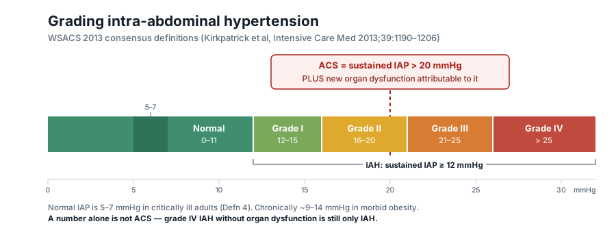
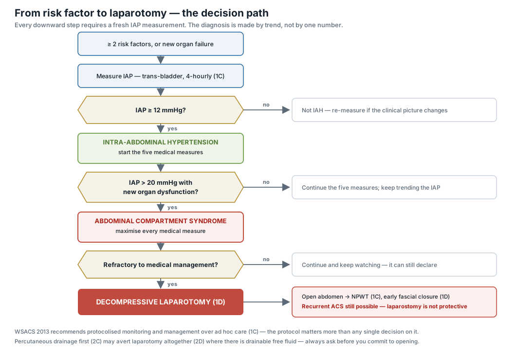
- Raised intra-abdominal pressure (intra-abdominal hypertension) is defined by a sustained or repeated pathological elevation in intra-abdominal pressure ≥ 12 mmHg
- Normal = IAP 5-7 mmHg
- Grade I = IAP 12–15 mmHg
- Grade II = IAP 16–20 mmHg
- Grade III = IAP 21–25 mmHg
- Grade IV = IAP >25 mmHg
- Abdominal compartment syndrome is defined as a sustained intra-abdominal pressure > 20 mmHg and new organ dysfunction that is attributable to elevated intra-abdominal pressure
- However, figuring out if organ failure is caused by abdominal compartment syndrome as opposed to other causes is subjective; the kidneys are quite sensitive to increased abdominal pressure (i.e. if urine output is adequate it’s unlikely the patient has abdominal compartment syndrome)
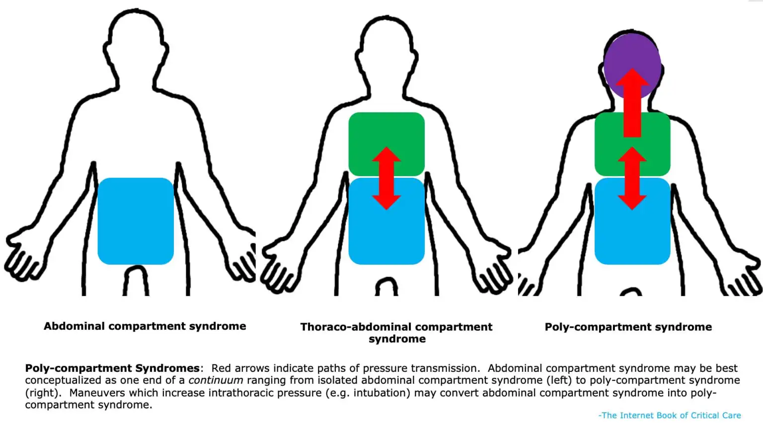
- Polycompartment syndrome is a condition where two or more anatomical compartments have elevated compartmental pressures
Risk Factors
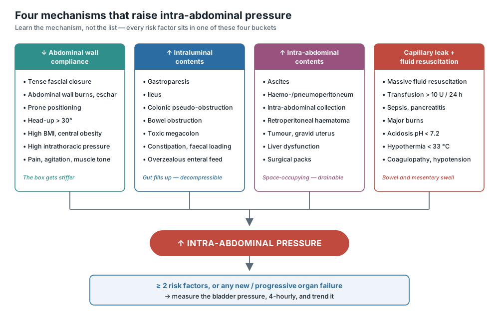
- Decreased abdominal wall compliance
- Acute respiratory failure with elevated intrathoracic pressure
- Abdominal surgery with fascial closure
- Major trauma/burns
- Prone positioning
- Head > 30°
- High BMI/central obesity
- Increased intra-luminal contents
- Gastroparesis
- Ileus
- Colonic pseudo-obstruction
- Increased abdominal contents
- Haemo/pneumoperitoneum
- Ascites
- Liver dysfunction
- Capillary leak or fluid resuscitation
- Acidosis (pH < 7.2)
- Hypotension
- Hypothermia (T < 33°C)
- Massive transfusion (> 10 U in 24 hrs)
- Coagulopathy
- Massive fluid resuscitation
- Pancreatitis
- Sepsis
- Oliguria
- Damage control laparotomy
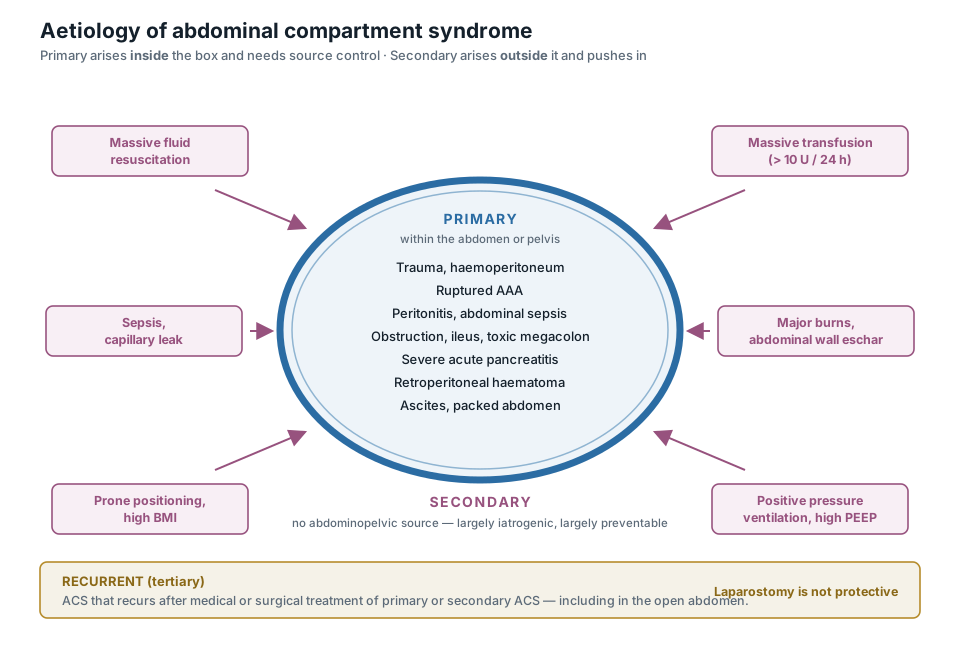
Aetiologies
| Primary | Secondary |
|---|---|
| Intra-abdominal infection | Massive transfusion |
| Abdominal trauma | Massive fluid resuscitation |
| Ascites | Sepsis |
| Ileus | Mechanical ventilation |
| Intra-abdominal haematoma | Abdominal wall burns |
| Laparoscopy with excessive inflation pressures | High body mass index |
| Acute Pancreatitis | Prone positioning |
| Peritoneal dialysis | |
| Dilated viscera (e.g. severe ileus, SBO, toxic megacolon) | |
 |
Complications
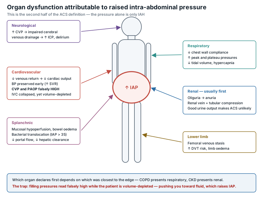
- Respiratory
- Decreased respiratory compliance ⇒ ↑ peak airway pressures → ↑ risk of atelactasis and pneumonia
- Decreased respiratory compliance ⇒ ↓ tidal volume
- Cardiovascular
- Decreased preload → decreased cardiac output
- Peripheral vascular resistance is elevated due to renal compression resultining in maintained systolic blood pressure
- Decreased preload → decreased cardiac output
- Gastrointestinal
- Direct compression
- Portal vein thrombosis
- Decreased gastric emptying
- Bowel wall ischaemia and oedema
- Translocation of bacterial products
- Increased intracranial pressure
- Renal failure
- Ileus
- Lower limb venous thrombosis
Clinical Features
- Initial manifestations vary depending on patient’s underlying physiology, for example:
- A patient with tenuous renal function could manifest with renal failure
- A patient with severe, chronic COPD might present with respiratory distress
- Palpation is only ~50% sensitive for abdominal compartment syndrome (i.e. abdomen can still be soft)
- However a tense abdomen is ~80% specific for abdominal compartment syndrome
- Abdominal pain and/or bloating are the commonly reported symptoms
- Other symptoms/signs are often a result of complications
- Oliguric acute renal failure is generally one of the earliest manifestations of abdominal compartment syndrome
- Increased work of breathing or increased airway pressures on the ventilator
- Raised ICP can also cause delirium or coma (occurs due to elevations of central venous pressure)
Management
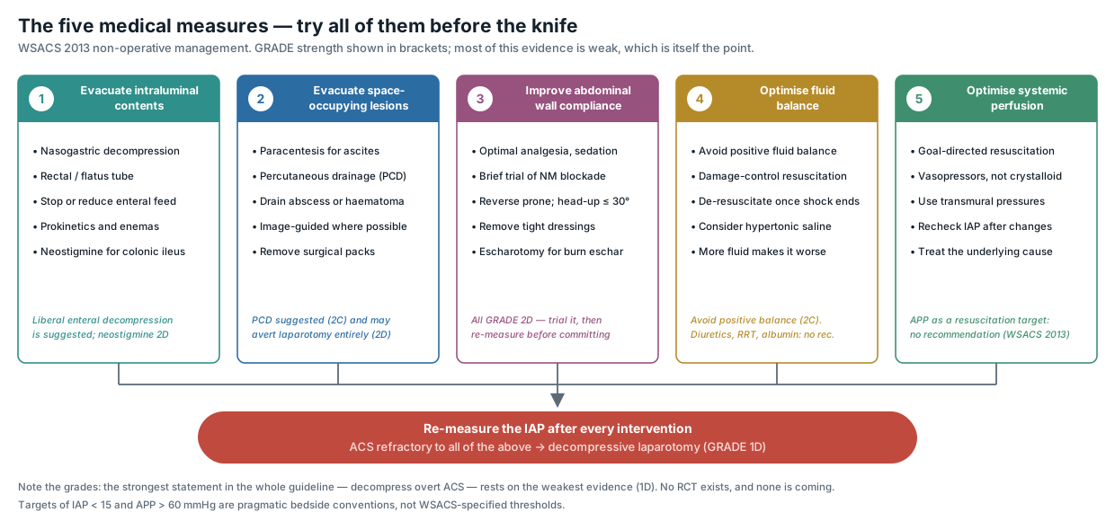
- Monitor intra-abdominal pressure when a patient has ≥2 risk factors or in the presence of new or progressive organ failure
- Prevention
- Controlled fluid resuscitation
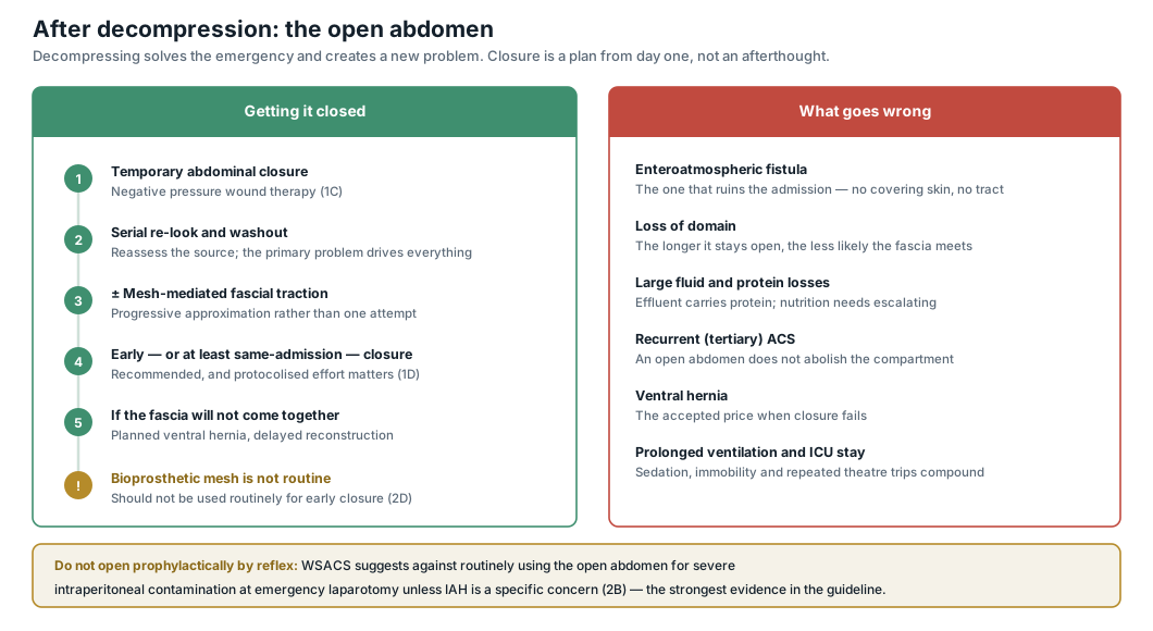
- Consider leaving the abdomen open, for staged closure
Haemodynamics
- Maintain a abdominal perfusion pressure > 60 mmHg with vasopressors (as additional crystalloids may merely aggravate abdominal tissue oedema)
- Use a to achieve this
- In established abdominal compartment syndrome, the patient is often intravascularly volume-depleted (despite tissue oedema), making it difficult to remove fluids without worsening haemodynamics
- Abdominal compartment syndrome may compress the inferior vena cava making it look empty
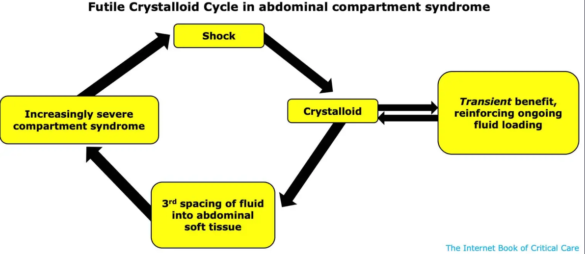
- Avoid fluid administration as while this may help temporarily, fluid rapidly transudates into the tissues which worsens swelling and increases intra-abdominal pressure
Decompress the Abdomen
- Evacuate Intra-Luminal Contents
- Nasogastric decompression
- Rectal decompression
- Prokinetics
- Minimise enteral nutrition
- Enemas
- Evacuate Abdominal Fluid Collections (e.g. paracentesis, percutaneous drainage, surgical evaluation)
- Surgical decompression with delayed closure
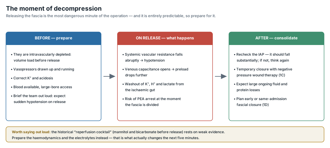
- On release be aware of hypotension (PEA arrest from reperfusion and a sudden decrease in systemic vascular resistance)
- Following fascial release, patients may experience severe ischaemia-reperfusion event, requiring haemodynamic support
- Post-operatively, patients will be left with an open abdomen which may be closed after 1-2 weeks following the resolution of the primary process
Decompress the Thorax
- Reduction of PEEP and plateau pressure on the ventilator may decrease intra-abdominal pressure
- Drainage of a large pleural effusion if present
- Intubation converts the thorax from a negative pressure system to a positive pressure system which may increase abdominal pressures, precipitating multi-organ failure with haemodynamic collapse
Sedation and Paralysis
- Sedation and paralysis may relax the muscles of the abdominal wall and improve abdominal compliance thereby reducing abdominal pressure
- However, in patients who are spontaneously breathing, their work of breathing decreases the intrathoracic pressures; eliminating this will transition to purely, positive pressure ventilation increasing their intrathoracic pressure and increase the intra-abdominal pressure
Other Measures
- Avoid head elevation > 30 degrees
- Remove constrictive dressing
- Eschars