- See Shock
Overview of RUSH Exam
- Heart: LV function, RV dilation, pericardial effusion/tamponade
- IVC: size and collapsibility (volume responsiveness)
- Lungs: B-lines (pulmonary oedema), pneumothorax (absence of sliding)
- Abdomen: free fluid (haemoperitoneum, ruptured AAA)
- Aorta: AAA
- Lower limbs: DVT (if PE suspected)
Can use the mnemonic HI-MAP (Heart, IVC, Morrison’s pouch, Aorta, Pneumothorax) or alternatively go in the approach of pump (heart), tank (IVC, Morison’s pouch), and pipes (aorta, DVT)
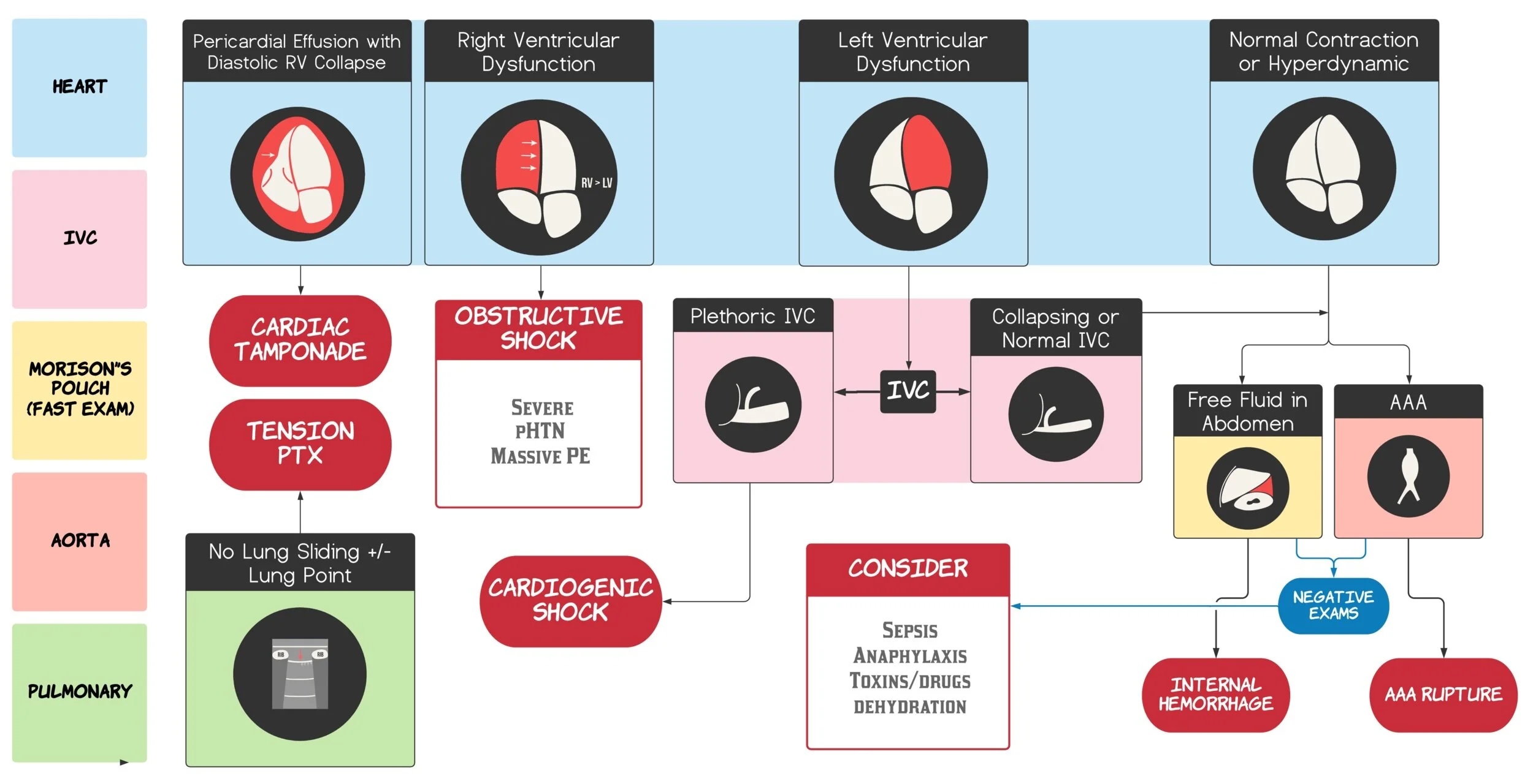
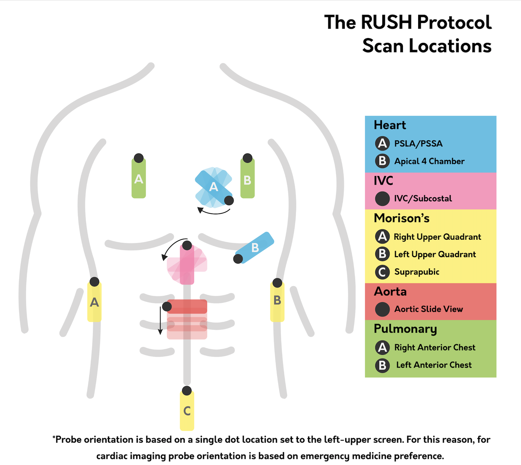
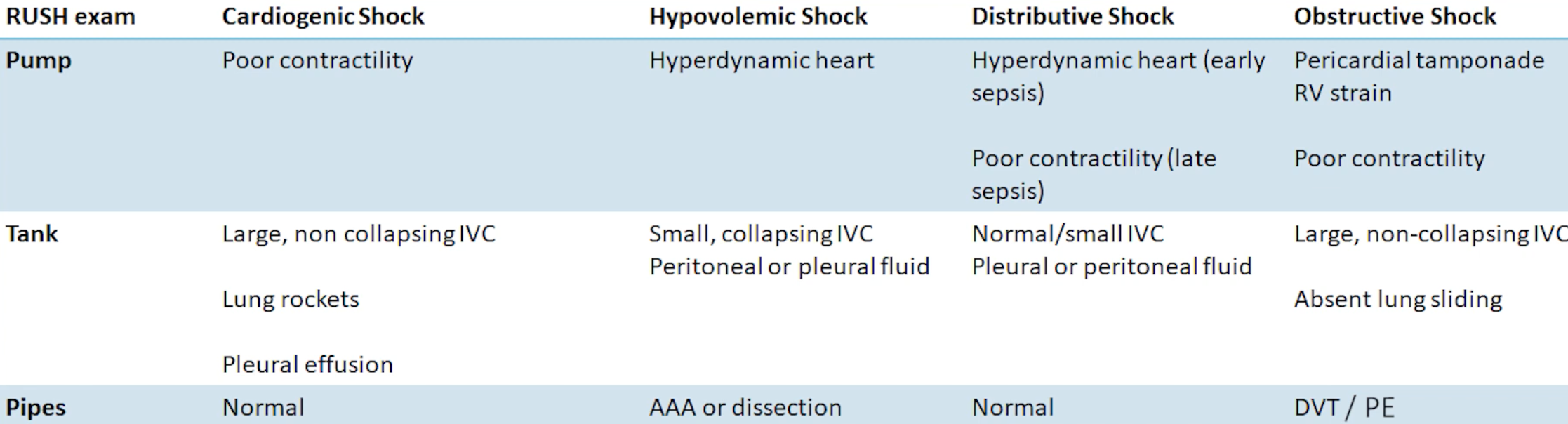
Heart
- Begin in the parasternal long axis view (see echo notes) and observe for:
- Pericardial effusion especially a circumfrential pericardial effusion
- Assessment of LV ejection fraction (can be just a visual qualitative assessment)
- Move to the parasternal short axis view and assess for:
- Right ventricular dilation/strain
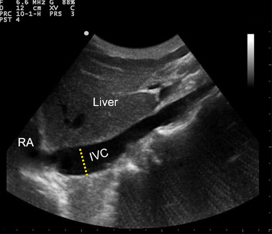
IVC view
- Review the IVC in the subcostal view with the probe marker pointing towards the patient’s head
- Qualtiative assessment of IVC:
- A flat IVC suggests distributive or hypovolaemic shock
Morrison’s Pouch
- Right upper quadrant view is most sensitive so often start at this
Aorta
- Look in the 3cm just above the umbilicus
- If the infra-renal abdominal aorta is >3cm and the patient is hypotensive, assume it is ruptured as POCUS is not sensitive enough for retroperitoneal bleed
Pneumothorax
-
Look at both anterior lung zones to observe for lung sliding (see Lung POCUS) or B-lines suggesting fluid overload
-
Bedside ultrasound
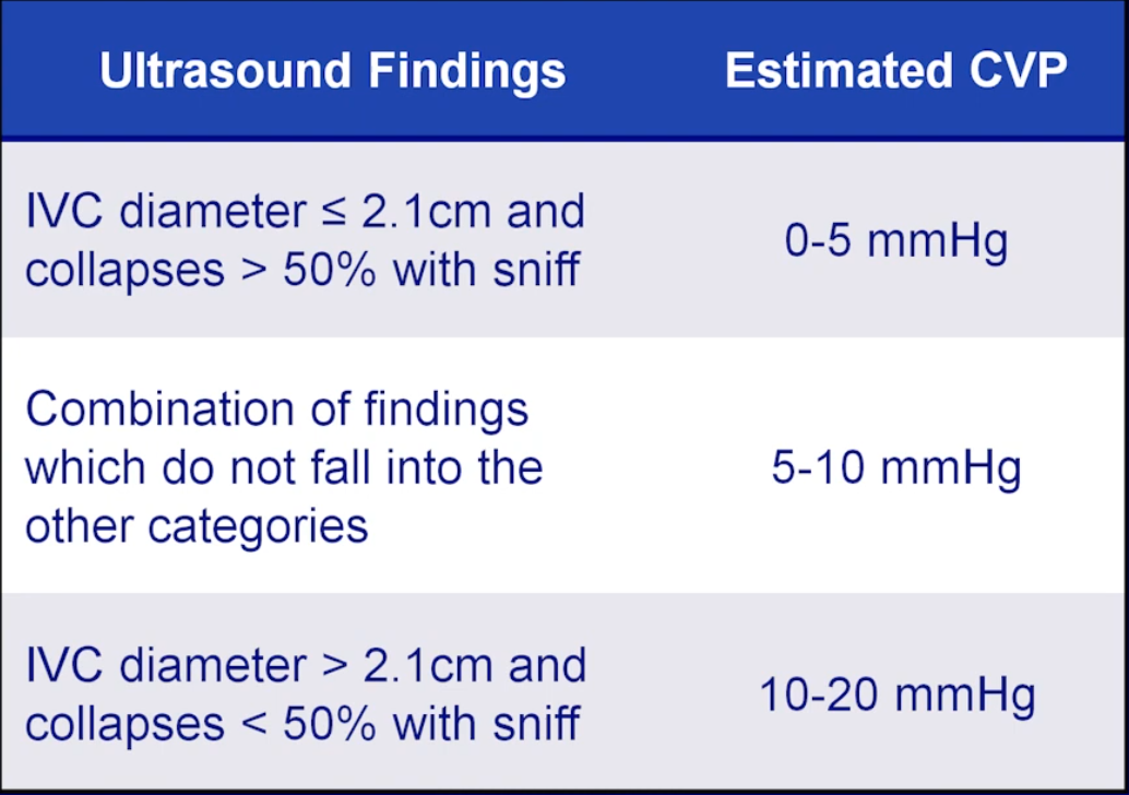
- Place in subxiphoid/subcostal area and orient head to toe
- Measure the IVC diameter and collapsibility with sharp inspiration (ask the patient to ‘sniff’)
-
Estimating CO
- Using LV function on ultrasound as a substitute for CO
- Use the ultrasound in the parasternal long axis view
Next Steps
- Once the aetiology of shock is found, to determine whether the patient’s hypotension would respond with fluids or pressors assess their Fluid Responsiveness with the LVOT VTI